About
Fertility Medicine
Early Pregnancy
Gynaecology
Treatment and Procedures
Clinic Info
Contact


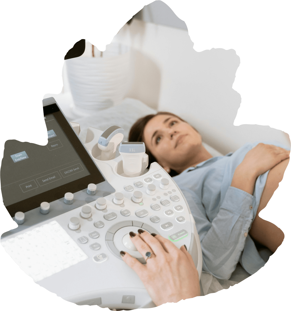
Some of these waves are reflected and are processed by the ultrasound machine to form pictures. These pictures are shown on a TV screen and recorded.
Ultrasound has been around for about 60 years now and studies have shown that it is a safe technique with no harmful side effects.
You will be shown into the ultrasound room by the nursing staff and asked to lie down on the examination chair. A jelly like substance is then placed on your skin or internally in the area of interest. The sound waves don’t travel through air so this allows transmission of the sound waves into your body. The probe produces sound waves that will form the images. You will be completely unaware of these sound waves and there should be no discomfort during the examination apart from a little pressure.
You may be asked to hold your breath – this is very important because when you breathe the organs go up and down in the abdomen. When you hold your breath the organs stay still allowing Dr Huang to get a better view of them.
The best technique for looking at the female pelvis is by performing an internal scan. This procedure is only performed with your consent and where appropriate for the area Dr Alice Huang is concerned about. Dr Huang will explain in detail what is involved. Remember, you are under no obligation to have this done although the ovaries etc are seen well and clearer images are taken. The sterilised probe (which is also covered by a protective sheath) is inserted into the vagina and manipulated very gently to show the anatomy in the pelvis.
It is a simple and short procedure every 5 years (previously 2 yearly) to check the health of your cervix. If the smear detects abnormal cellular changes or the presence of high risk human papillomavirus (HPV) is detected, then you will need a colposcopy.
A colposcopy is a magnifying scope that looks at the cervix to identify abnormal cells, to investigate abnormal cervical smears or cervical pathology. The cervix will be stained with acetic acid (vinegar) and iodine solution to show up abnormal cells. A biopsy (small piece of skin) may be taken of the abnormal part and sent away for pathological diagnosis. The examination can take around 25 minutes, and can feel like a prolonged cervical screening test. To facilitate thorough examination, you should not have your period at the time of colposcopy.
Contact us today to schedule a consultation and take the first step towards realising your parenthood goals.


